
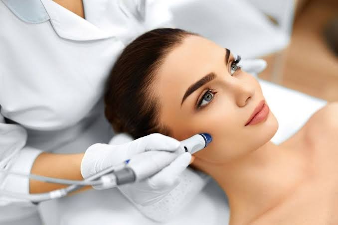
Laser energy represents one of the most thoroughly documented modalities in clinical aesthetics for reversing the visible markers of photoaging, persistent hyperpigmentation, and dermal scarring. When UV exposure, hormonal shifts, or past inflammation alter cutaneous architecture, conventional topical interventions often stall at the stratum corneum. Achieving true, long-lasting clinical clearance requires targeting subsurface chromophores—specifically melanin and hemoglobin—as well as the fibroblasts responsible for structural integrity. At Rania Aesthetic Studio, clinical protocols prioritize precision delivery, skin-type evaluation, and evidence-backed laser physics to restore smooth, luminous tone across diverse complexions.
Understanding Dermal Targets: Pigment, Scars, and Structural Decline
Hyperpigmentation develops when melanocytes produce an excess of pigment in response to chronic solar exposure, endocrine fluctuations, or injury. This manifests as discrete solar lentigines, diffuse melasma, or post-inflammatory hyperpigmentation. Because these melanin deposits reside across both epidermal and upper dermal layers, surface exfoliants rarely address the deeper source.
Acne scars and textural irregularities stem from abnormal wound healing. When severe inflammation damages native collagen networks, the skin produces disorganized fibrous tissue during repair, resulting in atrophic indentations such as boxcar, rolling, or ice-pick scars. Similarly, intrinsic aging paired with photo-damage leads to progressive elastosis, thinning epidermis, and degraded type I collagen bundles, forming static rhytids and skin laxity.
Clinical laser modalities resolve these conditions through selective photothermolysis. By matching a specific wavelength of light to a distinct target—melanin in dark spots, microvasculature in erythema, or water in surrounding tissue—the laser delivers rapid thermal energy. This breaks down excess pigment particles for cellular phagocytosis or creates microthermal treatment zones that prompt immediate neocollagenesis.
Comparing Laser Modalities for Corrective Outcomes
Different dermatological concerns demand distinct wavelengths, pulse durations, and delivery mechanisms to deliver optimal clearance without compromising tissue integrity.
- Q-Switched and Picosecond Lasers: Operating in nanosecond to picosecond domains, these systems generate acoustic shockwaves rather than thermal diffusion alone. They fragment stubborn dermal and epidermal melanin deposits into microscopic particles, allowing the lymphatic system to clear them naturally without injuring surrounding melanocytes.
- Fractional Non-Ablative Resurfacing: Utilizing wavelengths such as 1550 nm or 1927 nm, these systems create microscopic columns of thermal coagulative injury while leaving the stratum corneum intact. This triggers rapid tissue regeneration, reduces fine lines, and evens out dyschromia with minimal patient downtime.
- Fractional Ablative Systems: Using carbon dioxide (10,600 nm) or Erbium:YAG (2940 nm), these lasers vaporize targeted microscopic columns of tissue down to the reticular dermis. They remain the benchmark for deep atrophic scar remodeling and severe photodamage by forcing aggressive structural reorganization and long-term collagen synthesis.
- Intense Pulsed Light (IPL) and Broad-Band Light: While technically non-coherent light rather than a single laser wavelength, these multi-spectrum devices effectively sweep through superficial freckling, redness, and sun damage across larger surface areas.
What Real Recovery and True Results Look Like
Patients seeking laser correction must understand that clinical skin transformation follows biological healing timelines rather than instantaneous change.
- Pigment Clearing (Weeks 1 to 3): Superficial sun spots often darken into a “coffee ground” appearance immediately following therapy before naturally sloughing away within 7 to 10 days. Deeper dermal pigment gradually lightens over multiple sessions as macrophage clearance progresses.
- Scar Remodeling (Months 1 to 6): Because neocollagenesis and tissue remodeling are slow biochemical processes, scar depth reduction continues long after treatment sessions conclude. Real improvement emerges gradually as newly formed collagen lifts atrophic depressions.
- Texture and Firmness (Weeks 4 to 12): Fibroblast stimulation thickens the dermal matrix, yielding noticeable improvements in pore diameter, fine lines, and skin reflectivity within a few months of completed protocols.
Candidate safety remains paramount. Higher Fitzpatrick phototypes (IV through VI) carry an increased risk of post-inflammatory hyperpigmentation if excessive thermal spread occurs. Practitioners must tailor energy fluences, employ active epidermal cooling, and select appropriate wavelengths to ensure safe, predictable outcomes for every skin tone.
Sustaining Clinical Outcomes Through Expert Care
Laser resurfacing provides transformative correction, but long-term maintenance relies on diligent photoprotection and guided clinical aftercare. Exposure to broad-spectrum ultraviolet radiation without defense can rapidly reactivate underlying melanocytes and degrade newly deposited collagen. Daily broad-spectrum SPF 30 or higher, paired with physician-grade topical antioxidants, protects your investment and prevents recurrent dyschromia.
Selecting an experienced clinical provider ensures that your skin profile is meticulously mapped before an energy device ever touches your face. For clients pursuing medical-grade laser treatment in Sugarland, our experienced practitioners conduct thorough assessments to design customized, condition-specific treatment plans. Through careful wavelength calibration, advanced safety measures, and tailored aftercare, targeted laser therapy offers genuine, measurable restoration for clearer, firmer, and healthier skin.